

 EN

how-fiber-diameter-affects-energy-spread-during-endolaser-work, how-fiber-diameter-affects-energy-spread-during-endolaser-work, /blog
EN
The precision of energy delivery in minimally invasive procedures depends critically on understanding how fiber diameter influences light distribution. During endolaser work, the relationship between fiber dimensions and energy spread directly affects treatment efficacy, patient safety, and clinical outcomes across aesthetic and therapeutic applications.
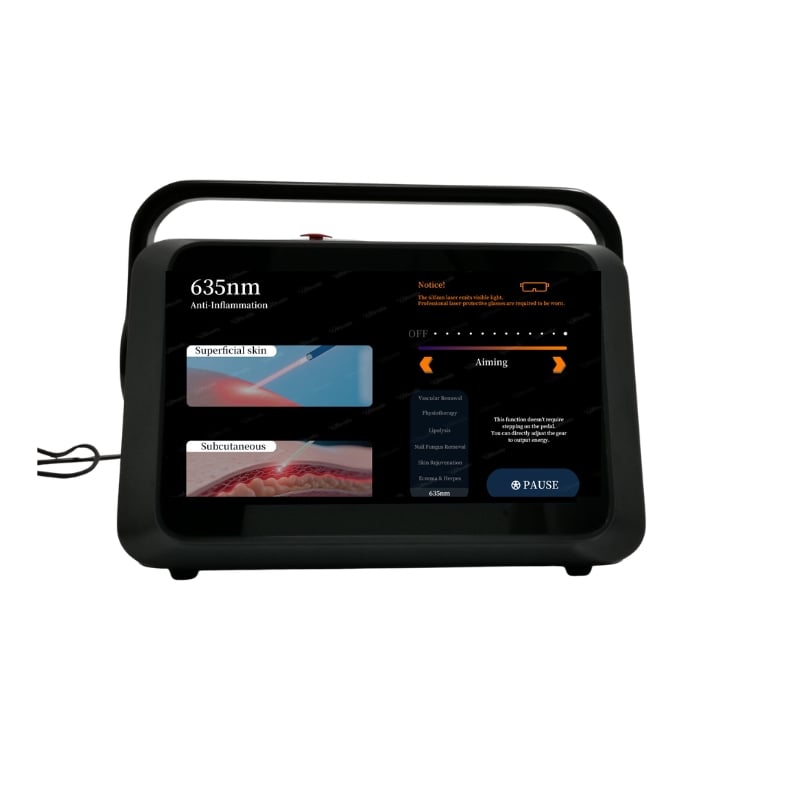
Medical professionals using laser lipolysis fiber and other minimally invasive laser device technologies must understand the physics governing light propagation through optical fibers. The core diameter, cladding thickness, and numerical aperture collectively determine how thermal energy disperses into surrounding tissue, influencing treatment depth, lateral spread, and thermal margin management during endolaser treatment procedures.
The core diameter of an optical fiber serving an endolaser system directly controls the initial beam width at the fiber tip. Smaller core diameters, typically ranging from 300 to 600 micrometers in clinical applications, create more focused energy delivery with sharper thermal gradients. Larger diameter fibers, measuring 900 micrometers to 1.5 millimeters, produce wider divergence angles and broader thermal zones. This fundamental principle governs how a minimally invasive laser device distributes photonic energy throughout target tissue.
When laser lipolysis fiber technology delivers energy through smaller cores, the resulting beam remains tightly collimated near the tip, then expands at a predictable rate determined by diffraction. Clinicians using endolaser treatment systems must recognize that fiber diameter selection directly impacts the thermal footprint—a 400-micrometer fiber creates a different energy distribution pattern than an 800-micrometer fiber delivering identical wattage. This distinction proves critical in sensitive anatomical locations where collateral thermal damage must be minimized.
Numerical aperture (NA), calculated from core diameter and cladding refractive index, determines the cone angle at which light exits an endolaser fiber tip. Higher NA values produce wider divergence angles—typically 25 to 35 degrees for clinical fibers—allowing energy to spread more rapidly into surrounding tissue. Lower NA configurations maintain tighter beam focus over extended distances, preserving lateral energy concentration. The laser lipolysis fiber specifications must balance NA to achieve desired penetration depth while controlling radial heat distribution.
In minimally invasive laser device applications, NA directly influences thermal gradient steepness. A fiber with 0.39 NA behaves distinctly differently than one with 0.22 NA, even when core diameter appears similar. Endolaser treatment protocols must account for these optical properties because they determine whether energy concentrates in a narrow thermal column or spreads diffusely through a wider zone. Practitioners must verify NA specifications when selecting endolaser systems for specific clinical indications.
Fiber diameter directly correlates with the depth at which an endolaser achieves maximum thermal effect. Smaller core diameters create sharper, more focused heating patterns that concentrate energy near the fiber tip, suitable for superficial or precisely targeted structures. Larger diameter fibers distribute energy more gradually, creating deeper penetration with gentler thermal gradients that may prove beneficial for diffuse therapeutic applications. The choice between fiber geometries fundamentally shapes clinical outcomes in endolaser treatment.
A minimally invasive laser device using a 400-micrometer laser lipolysis fiber deposits thermal energy within millimeters of the fiber track, generating precise tissue effects suitable for delicate procedures. Conversely, a 1-millimeter diameter endolaser fiber spreads energy across a broader thermal zone, potentially reaching deeper anatomical layers. Clinicians selecting equipment must understand how their chosen fiber diameter aligns with target tissue architecture and desired thermal depth to ensure safe, effective endolaser treatment delivery.
Energy spread during endolaser work creates thermal margins around the primary treatment zone that must remain within clinically acceptable limits. Smaller fiber diameters generate steep thermal gradients, meaning temperature drops rapidly beyond the primary thermal column—an advantage when protecting adjacent vital structures. Larger fibers produce gentler gradients, with thermal effects extending further laterally, requiring greater physical separation from sensitive tissues. A minimally invasive laser device selection must balance efficacy with collateral thermal protection.
During endolaser treatment procedures, practitioners must maintain awareness that fiber diameter selection affects the thermal safety margin. Vessels, nerves, or other structures near the treatment site experience different thermal stress depending on whether a small-diameter laser lipolysis fiber or large-diameter endolaser fiber performs the procedure. Smaller fibers generally offer superior precision in critical zones, while larger fibers may necessitate greater physical distance from vulnerable structures, influencing treatment planning and patient positioning.
Effective endolaser treatment requires deliberate fiber diameter selection aligned with specific clinical objectives. Cosmetic applications using laser lipolysis fiber benefit from smaller diameters (400-500 micrometers) that create precise thermal effects within target tissue while minimizing surrounding thermal injury. Therapeutic applications utilizing minimally invasive laser device technology for vascular or lymphatic work may employ intermediate sizes (600-800 micrometers) balancing penetration depth with acceptable collateral effects. Strategic fiber selection transforms an endolaser from a generic tool into a precisely tuned therapeutic instrument.
Clinicians implementing endolaser treatment protocols should establish procedure-specific fiber diameter guidelines based on anatomical depth, tissue type, and protection requirements. A protocol for facial endolaser treatment might specify 400-micrometer fiber for epidermal and superficial dermal work, while deeper muscular or subcutaneous applications employ 600-800-micrometer fibers. This structured approach ensures consistency, improves clinical outcomes, and supports evidence-based practice with minimally invasive laser device systems.
The relationship between power delivery and fiber diameter significantly impacts thermal outcome consistency. Smaller diameter fibers require lower wattage to achieve equivalent tissue temperatures compared to larger fibers, because energy concentrates in a narrower cross-sectional area. An endolaser treatment protocol using a 300-micrometer laser lipolysis fiber at 20 watts produces different thermal effects than the same fiber at 40 watts. Understanding this relationship prevents inadvertent thermal overdose and optimizes results across endolaser applications.
Minimally invasive laser device operators must resist assuming that larger power settings always produce superior results. Instead, practitioners should calibrate power settings to their selected fiber diameter, treatment duration, and tissue response targets. A well-designed endolaser treatment program establishes power-to-fiber-diameter ratios that consistently achieve desired clinical effects while respecting thermal safety margins. This evidence-based approach transforms fiber diameter from a forgotten specification into an active clinical parameter driving treatment quality.
Smaller fiber diameters create narrower, more focused thermal footprints with steep temperature gradients around the laser lipolysis fiber track, while larger diameters produce wider thermal zones with gentler gradients. The endolaser energy spreads differently depending on fiber core size; a 400-micrometer fiber concentrates heat more sharply than an 800-micrometer fiber at identical power, directly influencing whether a minimally invasive laser device achieves precise targeting or broader tissue effects during endolaser treatment.
Fiber diameter is a fixed physical characteristic that cannot be adjusted during treatment—practitioners must select the appropriate endolaser fiber before the procedure begins. If energy spread proves inadequate or excessive during laser lipolysis fiber delivery, the clinician must stop, change to a fiber with different diameter specifications, and resume treatment. Most minimally invasive laser device systems maintain multiple fiber diameters to support varied clinical scenarios, requiring practitioners to plan fiber selection based on anticipated treatment requirements during endolaser treatment procedures.
Smaller fiber diameters (300-500 micrometers) generally prove superior for sensitive areas because they concentrate endolaser energy in narrow thermal columns, minimizing collateral heat spread to adjacent vulnerable structures. Laser lipolysis fiber selection for delicate procedures prioritizes precision over penetration depth, making small-diameter options ideal for facial, periorbital, or neurovascular regions where a minimally invasive laser device must protect critical tissues. Practitioners should reserve larger diameter fibers for anatomically forgiving areas where broader thermal effects and deeper endolaser treatment reach prove clinically advantageous.