

 EN

understanding-focal-zone-geometry-in-focused-shockwave-therapy, understanding-focal-zone-geometry-in-focused-shockwave-therapy, /blog
EN
Focal zone geometry describes where acoustic energy concentrates, how wide that region is, and how it changes with depth. In focused shockwave therapy, understanding this geometry helps clinicians relate device settings to a target structure rather than treating depth as an isolated number. The central question is practical: where will the energy be concentrated in relation to the anatomy being assessed?
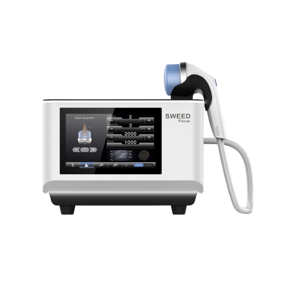
This matters because an extracorporeal shockwave is not distributed as a uniform beam through tissue. Its pressure field has a spatial shape, and the focused shockwave device, coupling method, treatment location, and patient anatomy all influence how the field meets the intended target. For orthopedic rehabilitation teams, focal-zone literacy supports clearer planning, more consistent communication, and sensible interpretation of treatment parameters.
Focal depth indicates the approximate distance from the applicator surface to the region where acoustic energy is most concentrated. In focused shockwave therapy, depth is a planning reference, not a guarantee that a target receives an identical dose in every patient. Tissue thickness, positioning, and the chosen coupling interface can affect the path from applicator to target, so clinicians should consider depth alongside anatomical assessment.
A focused shockwave device may offer adjustable focal depth, allowing the operator to align the energy concentration with structures at different distances from the skin. That flexibility is useful only when settings are matched to the clinical target. In orthopedic rehabilitation, a superficial region and a deeper attachment site call for different spatial considerations, even when the treatment area appears similar from the outside.
The focal region also has dimensions beyond depth. Its width and length describe the area in which energy is concentrated around the focal point, while energy outside that region does not simply disappear. This shape helps explain why focused shockwave therapy involves targeting and positioning rather than aiming at a broad area without reference to underlying anatomy.
When evaluating an extracorporeal shockwave system, clinicians can review how the manufacturer defines focal dimensions and what measurement conditions apply. A focused shockwave device specification should be interpreted as technical guidance, not as a stand-alone clinical outcome. In orthopedic rehabilitation, these dimensions help teams think through target coverage, applicator orientation, and whether a treatment plan needs careful repositioning across a larger region.
Treatment setup begins with identifying the structure of interest and estimating its location relative to the skin. Focused shockwave therapy then uses that assessment to guide applicator placement and the selection of a suitable depth setting, where available. Clinicians should account for patient position and landmarks, because a small change in angle or contact can alter the relationship between the focal region and the intended tissue.
An extracorporeal shockwave travels through a pathway, so the operator should also consider intervening anatomy and comfort. The focused shockwave device should be used according to its instructions, including specified coupling procedures and adjustment limits. For orthopedic rehabilitation, recording the selected location, patient position, and relevant settings can make follow-up sessions easier to interpret without assuming that identical numbers produce identical conditions in every case.
Orientation determines how the focal region sits in relation to the target. A focused shockwave device may be moved or angled to reach a clinically relevant location, but positioning should follow assessment rather than guesswork. Consistent contact and coupling help reduce avoidable variation at the skin interface, while clear communication with the patient supports a controlled, repeatable setup during focused shockwave therapy.
The treatment area may extend beyond the focal dimensions, particularly when the clinical plan addresses a broader region. In that situation, an extracorporeal shockwave protocol may involve deliberate repositioning rather than increasing exposure at one point without a reasoned plan. Orthopedic rehabilitation teams should document how positions relate to landmarks, helping distinguish a planned coverage pattern from unintentional movement between visits.
Device specifications can help explain focal depth and geometry, but they do not replace clinical judgment or establish which treatment is appropriate. Focused shockwave therapy decisions depend on assessment, treatment goals, contraindications, and the device instructions. A focused shockwave device parameter describes a technical condition; it should not be presented as a direct prediction of patient response or a guarantee of a particular result.
For an extracorporeal shockwave procedure, the relationship between settings and tissue response is affected by factors that a simple geometry diagram cannot capture. In orthopedic rehabilitation, clinicians can use specifications to support planning and explain the rationale for positioning, while monitoring tolerance and response in context. This approach keeps technical data useful without overstating what a focal-zone measurement can prove.
Useful documentation records the anatomical target, patient position, applicator orientation, selected depth or other relevant settings, and any repositioning during the session. Focused shockwave therapy notes become more interpretable when they distinguish intended focal placement from general treatment-area coverage. A focused shockwave device may present several adjustable parameters, so recording the settings that matter to the plan supports continuity across clinicians and visits.
Geometry is a framework for making setup more deliberate, not a substitute for training or patient-specific assessment. When an extracorporeal shockwave is delivered, the focal region should be considered together with anatomy, coupling, and the intended treatment objective. Orthopedic rehabilitation benefits from this practical view: teams can discuss why a location was chosen, what was adjusted, and how the session fits into the broader care plan.
Not exactly. Focal depth describes the location of the energy concentration under defined conditions, while actual treatment setup depends on anatomy, positioning, coupling, and device instructions. Focused shockwave therapy planning uses depth as one reference among several, rather than assuming the target will always sit at a precise distance.
Focal width helps describe the spatial extent of the concentrated region and can inform placement and coverage decisions. With an extracorporeal shockwave, it is useful to understand how the focal area relates to the target, while orthopedic rehabilitation planning still depends on the clinician’s assessment and the specific treatment objective.
No single setting should be assumed to fit every patient or target. A focused shockwave device setting must be selected within its instructions and considered alongside individual anatomy and clinical assessment. Focused shockwave therapy is planned around the intended target, and orthopedic rehabilitation teams should document relevant adjustments rather than rely on a universal preset.